Table of Contents (click to expand)
Odontogensis is the process of human teeth development. The formation of teeth takes place from tooth germ which is made of epithelial and mesenchymal tissue.
Many people identify their smile as the most attractive part of their persona, and what makes a good smile is some great pearly whites! We spend a great deal of time and money taking care of this public-facing body part. We brush our teeth twice a day, floss them (or at least we should), and bleach them, all in an effort to keep them shining.
However, have you ever taken a moment to think about how these bony structures erupt from our gums?

This unusual emergence strategy of teeth brought me to a rudimentary question… how do we grow teeth and where do they come from?
Recommended Video for you:
Initial Teeth Development
Humans have two sets of teeth: a deciduous set, also known as primary or baby teeth, and a permanent set, also referred to as secondary or adult teeth.
The formation of the deciduous dentitions begins before birth and erupts into the mouth soon after birth. Eventually, permanent teeth replace these milk teeth.
Odontogensis is the process of human teeth development. In utero, all the organs of your body develop from epithelial and mesenchyme tissues.
The formation of teeth takes place from the teeth germ, which is a clump of cells made of two basic components:
- epithelial tissue, derived from the ectoderm (the outermost layer of the germ cells of an embryo) and
- mesenchymal tissue, derived from the ectomesenchyme.
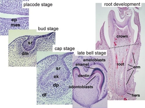
At around 5 weeks of gestational age, the epithelial layer thickens, forming a continuous band along the upper (maxilla) and lower (mandible) jaw. These horseshoe-shaped bands are known as the primary epithelial bands. These map out the position of the future dental arches of the jaw. Soon after, it grows into the underlying mesenchyme to form the dental lamina. Along the dental lamina, a series of epithelial clumps known as tooth germs form.
Specialized cells that form the mineralized portion of the tooth start differentiating during week six of gestational age.
Stages Of Tooth Development
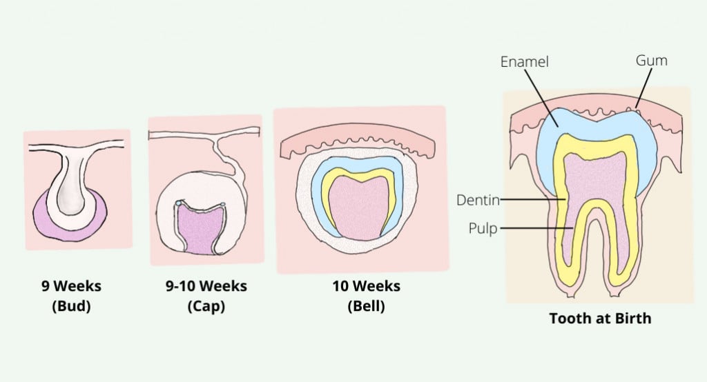
There are 3 stages of development of the tooth germ:
- Bud Stage
- Cap Stage
- Bell Stage
The process of tooth development comes to an end after the eruption of the tooth.
Bud Stage
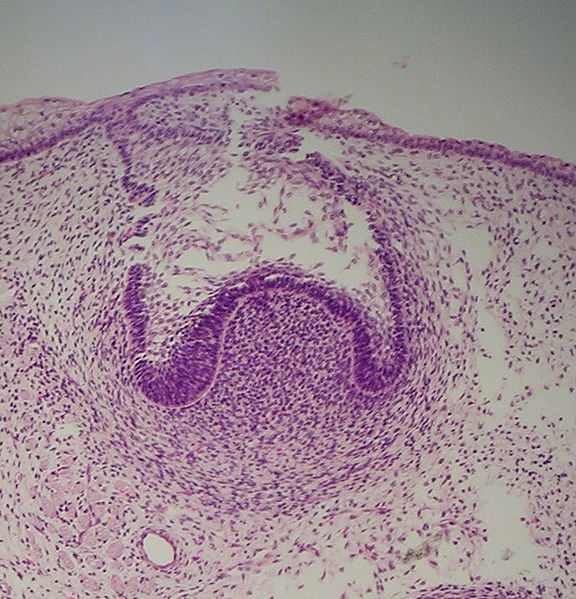
This is the first stage, and starts in week eight of gestational age. The first epithelial invasion into the underlying ectomesenchyme represents the beginning of the bud stage.
As the epithelial cells multiply into the ectomesenchyme, a swelling is formed adjacent to the epithelial bud. These swellings are called enamel organs. Each of the enamel organs later develops a tooth.
Cap Stage
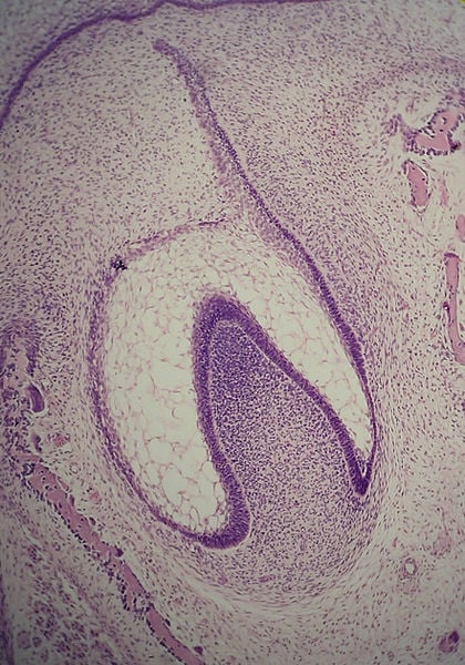
This stage is marked by the growth of the enamel organ. As the organ grows larger, it drags parts of the dental lamina along with it. The enlarging body resembles a cap sitting on the condensed ectomesenchyme, called the dental papilla. Thus, this stage of tooth development is known as the cap stage.
The enamel organ is surrounded by a fibrous capsule known as the dental follicle. It then develops into a periodontal ligament that anchors the root of the tooth to the bone. Hence, the enamel organ, dental papilla and dental follicle together serve as the tooth germ.
The center of the enamel organ forms a star-shaped layer and is therefore known as the stellate reticulum. The function of this layer is to maintain the shape of the tooth and protect the dental tissue lying beneath the layer.
Bell Stage
As the enamel organ continues to grow, it starts resembling a bell, so this stage is known as the bell stage.
During this stage, the tooth germ is ready for the formation of hard dental tissue. The cuboidal cells at the periphery of the enamel organ form the outer enamel epithelium, whereas the glycogen-rich columnar cells border the dental papilla from the inner enamel epithelium.
The cells at the periphery of the dental papilla initiate the formation of odontoblasts, which are dentine-secreting cells. Dentine is a mineral layer lying just beneath the enamel of the tooth. It is the supportive structure of the tooth, softer than the enamel, but harder than the bone.
The enamel formation sets off immediately after the first layer of dentine is laid down. Ameloblasts (enamel-secreting cells) are derived from the inner enamel epithelium. Enamel is the hardest layer of teeth and also the first line of defense that protects them from damage and decay. To know how corrosion of the enamel layer causes cavities, click here.
Upon completion of the enamel formation process, the ameloblast turns into the reduced enamel epithelium, which protects the enamel during the eruption. Root formation kicks off after the completion of crown formation.
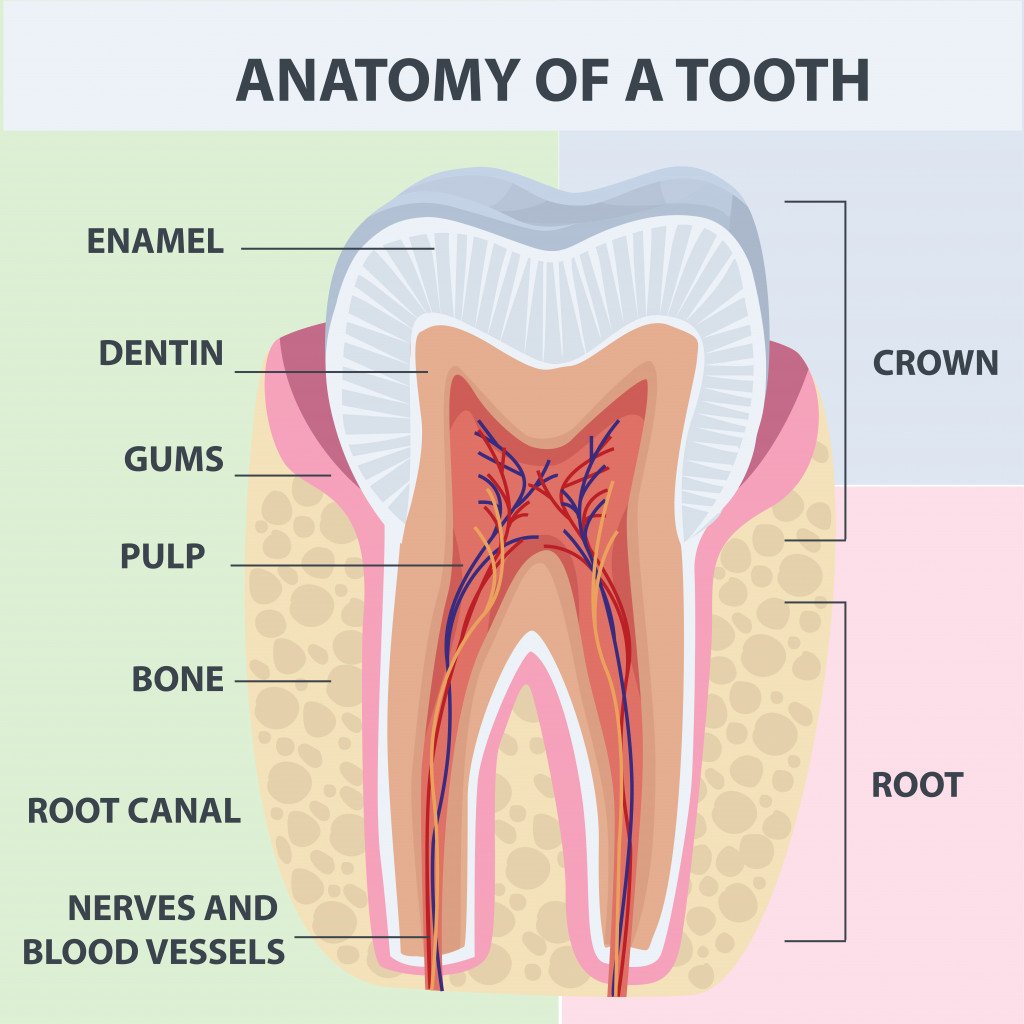
The cementum formation process, known as cementogenesis, occurs after the completion of the root. It is produced by cells known as cementoblasts. The cementum protects the root and acts as a surface for the periodontal ligament to anchor to the tooth.
As soon as the formation of the root begins, the tooth starts to erupt towards the surface of the gums. Thus, in humans, the first deciduous tooth starts pushing through the baby’s gum between the ages of 4 and 7 months.
At this point, the complex process of tooth formation comes to an end!
Conclusion
We embark on the journey of teeth formation just a few weeks into our embryonic life. A cluster of rudimentary cells differentiates to form tooth germs. These cells are capable of designing a robust mineral structure that allows us to perform the most important life-sustaining task of eating, thereby initiating the digestion process that supplies our body with essential nutrition!


