Table of Contents (click to expand)
The cerebrum is the largest part of the human brain. It is responsible for our voluntary functions and processes information from our sense organs. It develops prenatally, from the prosencephalon of the embryo. It is divided into 2 halves, the left and right hemisphere.
Humans are considered the most advanced species on the planet. We have the capacity to think, reason, apply logic, and so much more. Our emotional and logical capacity far surpasses that of other species. All the credit for this thinking ability goes to our brain, a tiny organ weighing about 3 pounds, which is what makes us so special.
This brain of ours is divided into 3 main parts – the cerebrum, cerebellum and brainstem. This article focuses on the first and largest part of the brain – the cerebrum.
Recommended Video for you:
Cerebrum Definition
The cerebrum is the largest part of the human brain. It is responsible for our voluntary functions and processes information from our sense organs. It develops prenatally, from the prosencephalon of the embryo. It is divided into 2 halves, the left and right hemisphere.
Cerebrum Structure
The cerebrum comprises the largest part of the brain. It develops prenatally, from the prosencephalon or the forebrain of the embryo. It is divided into 2 halves, the left and right hemisphere. The two hemispheres control opposite sides of the body, i.e., the right hemisphere controls the left side of the body and vice versa. The hemispheres are connected to each other by a structure called the corpus callosum, a dense, thick set of nerve fibers that divides the two hemispheres and facilitates communication between them.
The cerebrum can be divided based on different criteria which inform the way neuroscientists describe various brain structure. Here, we will look at the cerebral structure in two ways – the cerebral cortex and the cerebral hemispheres.
Grey And White Matter Of The Cerebrum
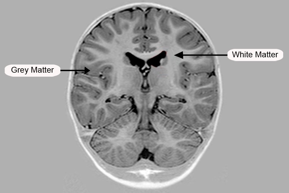
The cerebrum is divided into two parts, the outer grey matter covering the surface of the cerebrum called the cerebral cortex and the inner medullary region called the white matter.
The cerebral cortex which comes from the Latin word for bark is covered by a layer that is approximately 1/10th of an inch thick. This layer contains grooves and ridges, which increase the surface area of the cortex. There are about 20 billion neurons and over 300 trillion synapses in the cortex alone. An increased amount of surface area, therefore, allows for more neurons to be present.
One must have heard the term “grey cells” before, especially fans of Hercule Poirot. The fictional detective often referred to them as his, “little grey friends”. These refer to the cells in the cerebral cortex where the many neurons lack myelin sheath covering. The myelin sheath is a fatty covering around the axons of cells which give a whitish appearance to the inner medullary regions of the cerebrum.
On a microscopic look, the cerebral cortex contains six layers. These six layers have different neuron compositions and depending on the region and function of the cortical area, the size of the layers differ. Exceptions are the cortical area of the limbic system (in the temporal lobe) and the olfactory cortex which have only three layers.
Cerebral Lobes
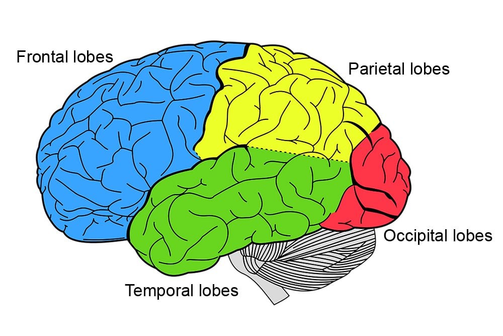
The cerebral hemispheres are divided into 4 different lobes. Starting from the forehead, the first lobe of the hemisphere is the frontal lobe, followed by the parietal lobe. The two are separated by the central sulcus (a sulcus is a groove or fissure). Below the frontal and parietal lobe is the temporal lobe. This is separated from the frontal lobe by the lateral sulcus. At the rear of the brain is the occipital lobe.
Delineating these brain areas helps neuroscientists pinpoint a certain brain region for study. It is also much easier to say that the temporal lobe rather than use confusing and lengthy anatomical directions to refer to it. These broad classifications of the brain help neuroscientists investigate the brain, and college students study it.
Besides broad lobes, neuroscientists have also developed more detailed maps of the brain. The Brodmann areas proposed by neurologist Korbinian Brodmann separated the cerebral cortex into 52 different areas based on the architecture of neurons in a region. Since 1909, when Brodmann first published his findings, neuroscientists have further subdivided these areas.
Cerebrum Functions
The cerebrum has many subdivisions, which control various functions. In a nutshell, the cerebrum controls all our voluntary functions, as well as our thinking, vision, hearing etc. Each of the lobes has its own specialized functions.
The frontal lobe handles our thinking, planning, short-term memory, etc. It is also a seat for most of the dopamine-delicate neurons. At the back of the frontal lobe is a section known as the motor area. This controls our voluntary actions. The left lobe also has a section called the Broca’s area, which is responsible for converting thoughts into words. Our parietal lobes control inputs like touch, temperature and taste, in addition to being responsible for our spatial balance and navigation.
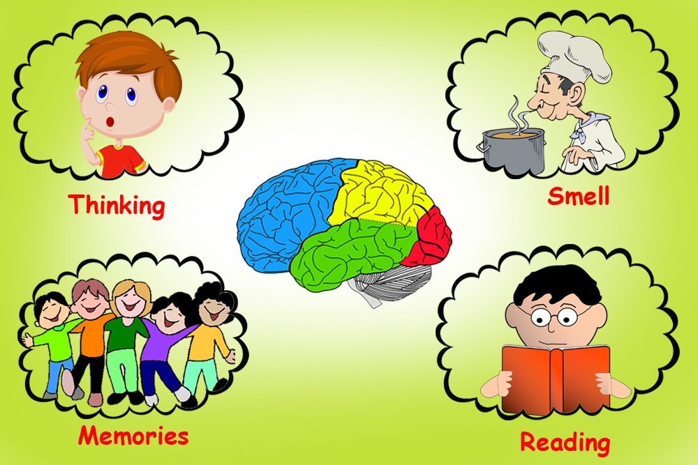
Our occipital lobe receives and processes all visual information, i.e., it is responsible for all our visual activity. Any damage to this area can lead to blindness. The temporal lobe takes care of inputs that elicit memories like visual or emotional memories.
Our cerebrum controls our thinking and cognitive functions, apart from our voluntary movements. Any damage to this area can result in serious consequences, such as paralysis, blindness, loss of hearing, emotional handicap, etc. Although the cerebrum doesn’t handle functions like heart rate or body balance, it can be safe to say that our cerebrum controls many of the critical functions that differentiate us from other species.

