Table of Contents (click to expand)
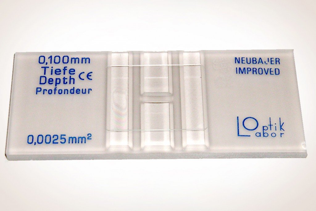
A hemocytometer is a specialized slide which is used for counting cells. It is actually a glass slide which has a 3×3 grid etched into it. Carved in it are intricate, laser-etched lines that form a grid. It also has its own coverslip.
Anyone who has anything to do with microbiology, biotechnology, pathology, or other related fields needs to be familiar with a hemocytometer. Used to count different microparticles or microorganisms, a hemocytometer is a special slide and much more expensive than an average glass slide.
It can be used to count the number of red blood cells in a sample and white blood cells, microbes such as yeast, and many others.
Recommended Video for you:
What Is A Hemocytometer?
The hemocytometer looks like an average glass slide, only heavier from a distance, but it is much more than that. It also has its own coverslip, which is different from a regular coverslip. On the slide, there are marked grooves that appear like an ‘H’. The horizontal line of the ‘H’ separates the 2 grids for counting. Therefore, each slide has two identical grids for counting cells. The depth of these 2 grids is a fixed 0.1mm
Let’s talk about the grids.
Hemocytometer Calculation
Each grid is a square with the dimensions of 3×3 mm2. This square has three equidistant vertical and horizontal lines. These divide it into 9 smaller squares of 1×1 mm2 each. These are separated from each other by triple-ruled lines. Of these 9 squares, the 4 corner squares are used to count bigger cells, like WBCs, while the center square is used to count smaller cells, such as RBCs.
The 4 corner squares of the main grid are further divided into 16 smaller cells. The center square of the main grid is divided into 25 smaller squares, each of which is again divided into 16 smaller squares. The sample to be counted is loaded onto the slide after the coverslip has been placed. Excess fluid drains into the grooves on the side. However, the person loading the sample must be extremely careful while loading.
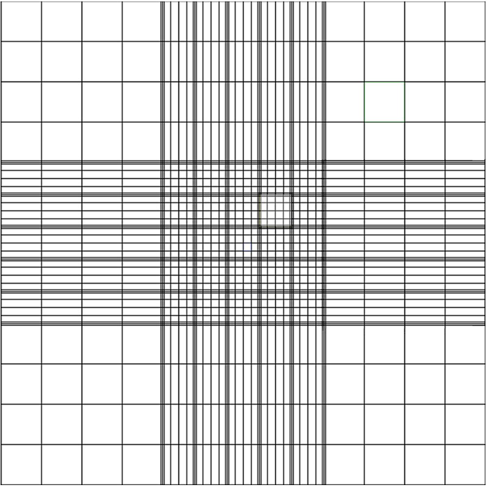
This covers the structure and design of the hemocytometer, but to understand how counting and calculation is done, let’s consider the example of counting WBCs for the corner squares, and RBCs for the center square.
Hemocytometer Counting
RBCs, being smaller in size and larger in number, are counted in the center square. This has a greater number of divisions and therefore makes counting easier. As mentioned above, the center square contains 25 smaller squares. The area of each of these is 1/25 mm2, which is 0.04 mm2. Once the sample is loaded, not all the cells are counted. Out of 25, any 5 squares are picked for the counting. The division of each of these 0.04 mm2 squares into 16 smaller ones makes it easier for the person to count the number of cells rather than just having to count in an empty square. There are a number of patterns to select the 5 squares that should be counted. The corner 4 and center square can be picked, or any of the diagonal lines of squares.
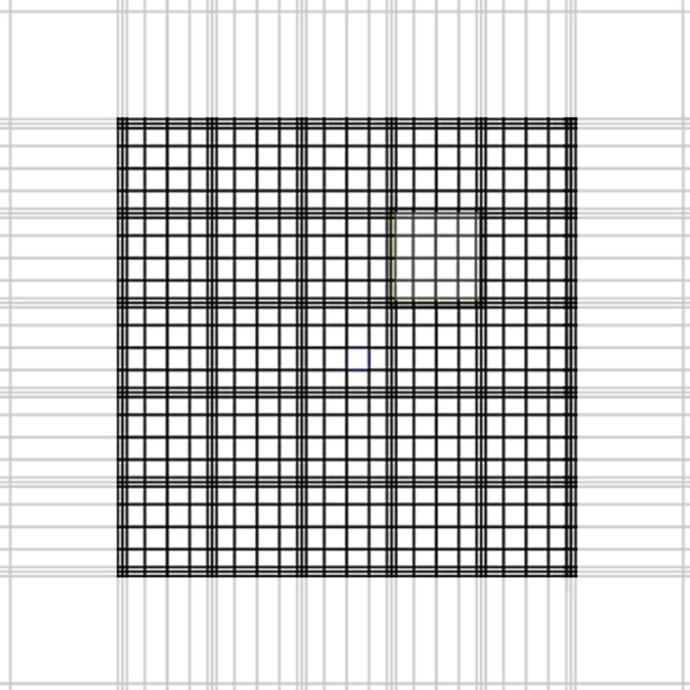
Once the number of cells in 5 squares has been counted, their mean is taken.
Let ‘n’ be the mean.
Therefore, the average number of cells in each of the tiny 0.04 mm2 squares is n. The volume of each of these cells is 0.04 x 0.1 = 0.004 mm3. The number of cells in 1 mm3 is n/0.004.
Thus, the total number of cells in 1ml is (n/0.004) x 1000. We multiply by one thousand as 1000 mm3 = 1 cm3; and 1 cm3 = 1 mL

WBC Count
When WBCs are counted, the calculation is much easier. WBCs are counted in the 4 corner squares of the main grid. These squares have an area of 1 mm2 each. To get the WBC count, the number of cells in each square are counted, and their mean is then calculated. Let the mean be ‘n’. The volume of each square is 1 x 0.1 = 0.1 mm3. The number of cells in 1 mm3 is n/0.1. Therefore, the total number of cells in 1ml is (n/0.1) x 1000. We multiply by one thousand as 1000 mm3 = 1 cm3; and 1 cm3 = 1 mL
Rules To Be Observed
While counting cells, certain things require attention. Some cells may not lie either inside or outside the square. Rather, they may fall on the border. Therefore, a simple practice of including cells that fall on the top and left border and excluding cells that fall on the bottom and right border is followed. However, this is not a rule. The basic principle is that any 2 adjacent borders should be counted, and the remaining 2 borders should be rejected. Also, this selection criteria must apply to all the squares being counted.
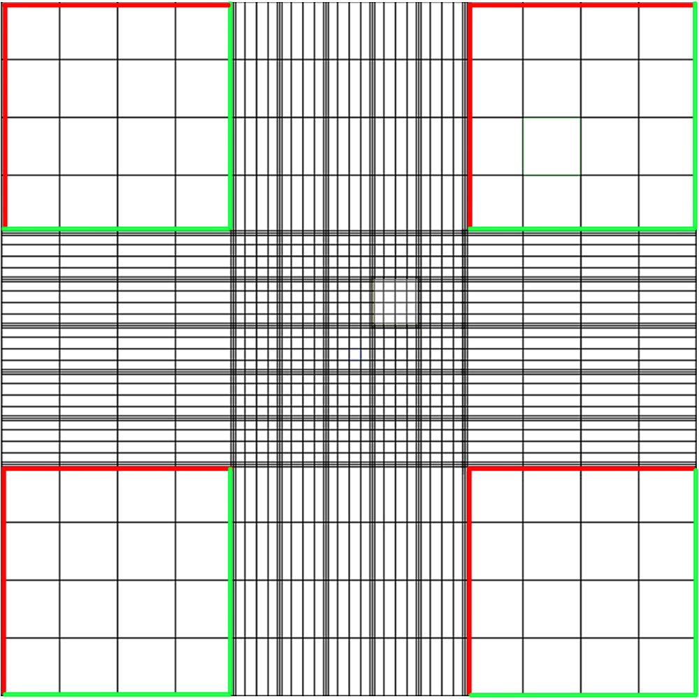
Sometimes the solution of the sample can be too concentrated. If it is too highly concentrated, the cells overlap and the counting is therefore incorrect.
Therefore, such concentrated cell solutions must be diluted with a suitable solution. This dilution must also be factored in the calculations. If, for example, the sample was diluted 10 times, the final answer from the calculations must be multiplied by 10.
Understanding how a hemocytometer work is necessary for a number of laboratory tests as they have an accuracy of within 20% of the automated answers.
So… you are welcome! You now know how to use a hemocytometer… theoretically.


